Overview
Source: Laboratory of Dr. B. Jill Venton - University of Virginia
Ultraviolet-visible (UV-Vis) spectroscopy is one of the most popular analytical techniques because it is very versatile and able to detect nearly every molecule. With UV-Vis spectroscopy, the UV-Vis light is passed through a sample and the transmittance of light by a sample is measured. From the transmittance (T), the absorbance can be calculated as A=-log (T). An absorbance spectrum is obtained that shows the absorbance of a compound at different wavelengths. The amount of absorbance at any wavelength is due to the chemical structure of the molecule.
UV-Vis can be used in a qualitative manner, to identify functional groups or confirm the identity of a compound by matching the absorbance spectrum. It can also be used in a quantitative manner, as concentration of the analyte is related to the absorbance using Beer's Law. UV-Vis spectroscopy is used to quantify the amount of DNA or protein in a sample, for water analysis, and as a detector for many types of chromatography. Kinetics of chemical reactions are also measured with UV-Vis spectroscopy by taking repeated UV-Vis measurements over time. UV-Vis measurements are generally taken with a spectrophotometer. UV-Vis is also a very popular detector for other analytical techniques, such as chromatography, because it can detect many compounds.
Typically, UV-Vis is not the most sensitive spectroscopy technique, because not a lot of light is absorbed over a short path length. Other spectroscopy techniques such as fluorescence have higher sensitivity, but they are not as generally applicable, as most molecules are not fluorescent. UV-Vis has a similar sensitivity to other absorbance measurements, such as infrared spectroscopy.
Principles
UV-Vis is often called a general technique because most molecules will absorb in the UV-Vis wavelength range. The UV extends from 100–400 nm and the visible spectrum from 400–700 nm. The 100–200 nm range is called the deep UV. Light sources are more difficult to find for this range, so it is not routinely used for UV-Vis measurements. Typical UV-Vis spectrometers use a deuterium lamp for the UV that produces light from 170–375 nm and a tungsten filament lamp for visible, which produces light from 350–2,500 nm.
When a photon hits a molecule and is absorbed, the molecule is promoted into a more excited energetic state. UV-visible light has enough energy to promote electrons to a higher electronic state, from the highest occupied molecular orbital (HOMO) to the lowest unoccupied molecular orbital (LUMO). The energy difference between the HOMO and the LUMO is called the band gap. Typically, these orbitals are called bonding and anti-bonding. The energy of the photon must exactly match the band gap for the photon to be absorbed. Thus, molecules with different chemical structures have different energy band gaps and different absorption spectra. The most common transitions that fall in the UV-Vis range are π-π* and n- π*. Pi orbitals arise due to double bonds, and n orbitals are for non-bonding electrons. Pi star are anti-bonding pi orbitals. Thus, the best UV-Vis absorption is by molecules that contain double bonds. Pi orbitals adjacent to each other that are connected, called conjugation, typically increases absorption. Sigma-σ* transitions, associated with single bonds, are higher energy and fall in the deep UV, so they are less useful for routine use. The appearance of broad bands or shoulders on the UV-Vis structure is due to the numerous vibrational and rotational states of a molecule, which lead to separate energy band gaps of slightly different energies.
For molecules with absorption in the visible region, the compounds will often appear colored. However, a common misconception is that the wavelength of peak absorption (λmax) for a compound is the color it appears. A compound that appears red does not have much absorption in the red region of the spectrum. Instead, the λmax for a compound that looks red is green. The color of a compound arises because those wavelengths of light are selectively transmitted through the sample, and thus they are not absorbed. A color wheel is helpful in determining what color a compound will absorb and what range the λmax will be, as the color directly across the wheel from the observed color is the color that is most absorbed.
Absorption follows Beer's Law, A=εbC where ε is the molar attenuation coefficient, b is path length, and C is concentration. The molar attenuation coefficient is the characteristic of an individual compound to absorb at a given wavelength and this property is due to functional groups, conjugation, etc. If a compound does not have a high attenuation coefficient, it could be tagged with an appropriate group to increase its absorbance. Path length is generally related to the size of the cuvette and is 1 cm in standard spectrophotometers.
UV-Vis is performed on a variety of instruments, from traditional spectrophotometers to more modern-day plate readers. The absorbance wavelength must be chosen, either using a filter or a monochromator. A monochromator is a device that separates the wavelengths of light spatially and then places an exit slit where the desired wavelength of light is. Monochromators can be scanned to provide a whole absorbance spectrum. Alternatively, a diode-array instrument allows all colors of light to be transmitted through the sample, then the light is separated into different wavelengths spatially and detected using photodiodes. Diode-array instruments collect full spectra faster, but are more complicated and more expensive.
Procedure
1. Calibrate the Spectrometer
- Turn on the UV-Vis spectrometer and allow the lamps to warm up for an appropriate period of time (around 20 min) to stabilize them.
- Fill a cuvette with the solvent for the sample and make sure the outside is clean. This will serve as a blank and help account for light losses due to scattering or absorption by the solvent.
- Place the cuvette in the spectrometer. Make sure to align the cuvette properly, as often the cuvette has two sides, which are meant for handling (may be grooved) and are not meant to shine light through.
- Take a reading for the blank. The absorbance should be minimal, but any absorbance should be subtracted out from future samples. Some instruments might store the blank data and perform the subtraction automatically.
2. Perform an Absorbance Spectrum
- Fill the cuvette with the sample. To make sure the transfer is quantitative, rinse the cuvette twice with the sample and then fill it about ¾ full. Make sure the outside is clean of any fingerprints, etc.
- Place the cuvette in the spectrometer in the correct direction.
- Cover the cuvette to prevent any ambient light.
- Collect an absorbance spectrum by allowing the instrument to scan through different wavelengths and collect the absorbance. The wavelength range can be set with information about the specific sample, but a range of 200–800 nm is standard. A diode-array instrument is able to collect an entire absorbance spectrum in one run.
- From the collected absorbance spectrum, determine the absorbance maximum (λmax). Repeat the collection of spectra to get an estimate of error in λmax.
- To make a calibration curve, collect the UV-Vis spectrum of a variety of different concentration samples. Spectrometers are often limited in linear range and will not be able to measure an absorbance value greater than 1.5. If the absorbance values for the sample are outside the instrument's linear range, dilute the sample to get the values within the linear range.
3. Kinetics Experiments with UV-Vis Spectroscopy
- UV-Vis can be used for kinetics experiments by examining the change in absorbance over time. For a kinetics experiment, take an initial reading of the sample.
- Quickly add the reagent to start the chemical reaction.
- Stir it well to mix with the sample. If a small amount is added, this could be done in a cuvette. Alternatively, mix the reagent with sample and quickly pour some in a cuvette for a measurement.
- Measure the absorbance at the λmax for the analyte of interest over time. If using up the reagent being measuring (i.e. absorbance is going up because there is less reagent to absorb), then the decay will indicate the order of the reaction.
- Using a calibration curve, make a plot of analyte concentration vs time, converting the absorbance value into concentration. From there, this graph can be fit with appropriate equations to determine the reaction rate constants.
Results
UV-Vis can be used to obtain a spectrum of colored compounds. In Figure 1A, the absorbance spectrum of a blue dye is shown. The background shows the colors of light in the visible spectrum. The blue dye has a λmax absorbance in the orange/red. Figure 1B shows a spectrum of a red dye, with λmax in the green.
Kinetics can be measured from a plot of absorbance at one wavelength over time. Figure 2 shows a plot of the absorbance of a blue dye (at 630 nm) as it reacts with bleach.
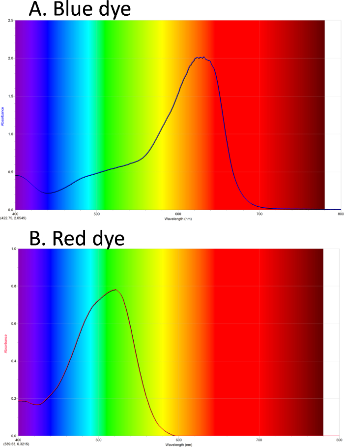
Figure 1. UV-Vis absorbance spectra. A. Blue dye #1 has maximum absorbance in the orange/red. B. Red dye #40 has maximum absorbance in the green. Please click here to view a larger version of this figure.

Figure 2. UV-Vis for kinetics. Absorbance of blue dye #1 as it reacts with bleach. The curve can be fit with an exponential decay, indicating first order kinetics. Please click here to view a larger version of this figure.
Applications and Summary
UV-Vis is used in many chemical analyses. It is used to quantitate the amount of protein in a solution, as most proteins absorb strongly at 280 nm. Figure 3 shows an example spectra of cytochrome C, which has a high absorbance at 280 and also at 450 because of a heme group. UV-Vis is also used as a standard technique to quantify the amount of DNA in a sample, as all the bases absorb strongly at 260 nm. RNA and proteins also absorb at 260 nm, so absorbance at other wavelengths can be measured to check for interferences. Specifically, proteins absorb strongly at 280 nm, so the ratio of absorbance at 280/260 can give a measure of the ratio of protein to DNA in a sample.
Most simple analyses measure the absorbance one wavelength at a time. However, more chemical information is present if measurements are made at many wavelengths simultaneously. Diode-array instruments capture all the light that is transmitted, split the light into different colors using a prism or holographic grating, and then absorbance at different wavelengths is captured on a linear array of photodiodes. The advantage of this method is that it is useful for measuring many different molecules simultaneously.

Figure 3. UV-Vis spectrum of a protein. The peak at 280 nm is indicative of a protein. The peak at 450 is due to absorbance of the heme group in cytochrome C.